


Advancing Precision in Breast Imaging
Early breast cancer detection relies on accurate imaging, yet not all breast types present the same diagnostic challenges. Women with smaller breast volumes often require specialized imaging techniques to ensure that subtle abnormalities are not overlooked. As part of our commitment to elevating breast health standards, we emphasize tailored approaches that enhance diagnostic clarity, improve patient comfort, and support timely detection. At Velox Imaging, our focus is on precision-driven methods that adjust to each patient’s unique anatomy.
Understanding the Complexity of Imaging Small Breasts
Breast size significantly affects image quality and the ease with which abnormalities can be detected. Smaller breasts have less fatty tissue, making dense glandular structures more prominent. While this may appear advantageous, high density can obscure lesions and reduce the clarity of standard images. To address this, specialized mammography techniques ensure consistent compression, enhanced resolution, and minimized tissue overlap.
Because every patient’s anatomy is unique, adapting imaging approaches helps radiologists gain clearer visibility. This is particularly important when performing mammography for small breasts, where capturing fine details is essential for a reliable diagnosis.
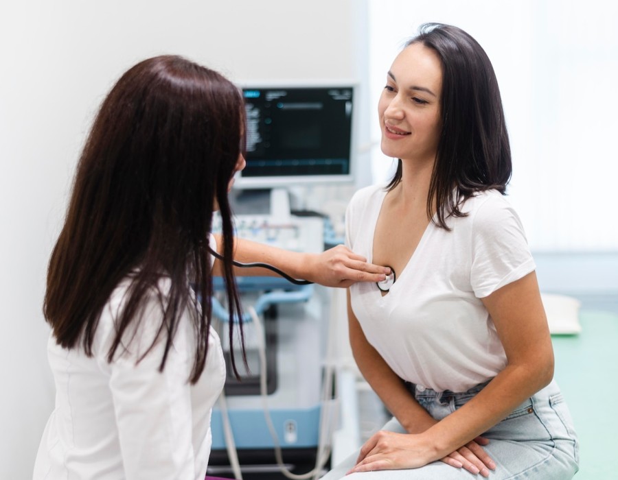
Why Standard Mammography May Not Be Enough
Traditional mammography equipment is designed for a broad range of body types, but it often requires modifications for individuals with lower breast volume. Without these adjustments, there is a greater risk of insufficient compression, imaging artifacts, or incomplete visualization of breast tissue. These limitations may lead to delayed diagnoses or unnecessary follow-up exams.
Facilities that specialize in breast mammography radiology understand the need for individualized imaging settings, adjusted paddle sizes, and enhanced software that compensates for anatomical differences. Such precision safeguards accuracy and ensures that no part of the breast is overlooked.
Specialized Techniques Used for Imaging Small Breasts
1. Customized Compression for Enhanced Accuracy
Compression is essential for clear, high-contrast mammographic images. With smaller breasts, achieving optimal compression requires time, precision, and specialized paddles. Proper compression spreads out glandular tissue, reduces motion artifacts, and improves visualization of microcalcifications - critical markers in early breast cancer detection.
Radiologists trained in breast mammography radiology utilize adaptive compression tools to ensure that imaging is both comfortable and diagnostically effective.
2. High-Resolution Digital Mammography
Digital technology elevates breast imaging quality, especially for patients with minimal breast tissue. High-resolution detectors capture fine details, enabling radiologists to differentiate between normal structures and suspicious findings more effectively. This is particularly important for mammography for small breasts, where small areas of concern must be magnified for precise evaluation.
Advanced digital systems also reduce noise and maintain consistent clarity across all breast sizes.
3. Tomosynthesis (3D Mammography) for Layered Clarity
3D mammography has transformed early breast cancer detection. By taking multiple images from different angles, tomosynthesis creates a layered reconstruction of breast tissue. This minimizes tissue overlap, which is a common challenge in small, dense breasts.
For women undergoing mammography for small breasts, 3D imaging significantly improves detection rates of tiny lesions and reduces false positives. It is especially valuable in identifying tumors hidden behind overlapping tissue structures.
4. Ultrasound and MRI as Complementary Tools
In scenarios where mammograms alone cannot provide complete clarity, supplemental imaging modalities are essential. Ultrasound enhances visualization of dense glandular structures, while MRI offers exceptional sensitivity for detecting early cancers. These supplemental tools ensure that smaller breast sizes do not compromise diagnostic accuracy.
Patients receiving care in advanced diagnostic imaging centers in Ontario benefit from multi-modal imaging options that enrich diagnostic outcomes.
Patient Comfort and Confidence in the Imaging Process
For many women, breast imaging can be intimidating - especially when prior experiences with inadequate imaging have resulted in repeat exams or inconclusive results. Providing comfort and reassurance during mammograms is essential to reducing anxiety and improving patient cooperation.
At Velox Imaging, we take a patient-centered approach by using ergonomically designed paddles, clear communication, and gentle positioning techniques. These elements create a positive experience while maintaining the highest imaging standards.
How Specialized Imaging Improves Early Detection
Enhanced imaging techniques directly contribute to improved early detection rates. With smaller breasts, there is less tissue to conceal abnormalities, but without proper imaging methods, subtle lesions may still be missed. Precision-based imaging ensures that every area of breast tissue is thoroughly evaluated.
When patients choose advanced diagnostic imaging centers in Ontario, they receive the benefit of equipment designed to optimize imaging for all breast sizes, as well as radiologists trained to interpret the nuanced differences in smaller breast anatomy.
Why Choosing the Right Imaging Facility Matters
Selecting a facility that prioritizes tailored imaging solutions is essential for accurate, reliable breast health assessments. Centers equipped with advanced technology, specialized paddles, tomosynthesis platforms, and experienced radiologists can ensure superior outcomes.
This is why many individuals trust Velox Imaging for their breast screening needs. Our focus on innovation, precision, and patient comfort allows us to deliver confidence-driven care that supports long-term health and peace of mind.
Conclusion: Advancing Breast Health Through Specialized Imaging
Effective breast cancer detection begins with imaging that adapts to each patient’s anatomy. Specialized techniques designed for small breast volumes ensure clarity, accuracy, and early detection capabilities that standard equipment cannot always achieve. With advancements in digital mammography, tomosynthesis, and supplemental modalities, women now have access to high-precision imaging tailored to their needs.
Facilities offering expertise in breast mammography radiology, and advanced technologies continue to elevate the standard of care. By choosing trusted providers in diagnostic imaging centers in Ontario, patients can be confident that every aspect of their breast health is evaluated with accuracy and care.
Comments