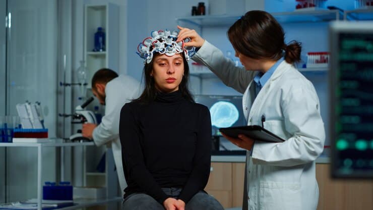
Astrocytomas are a group of brain tumors that develop from astrocytes, which are supportive cells within the central nervous system. These tumors vary in size, location, and aggressiveness. Because of these differences, astrocytoma treatment requires careful evaluation and a highly specialized approach. Neurosurgeons play a central role in diagnosing, planning, and performing surgical procedures aimed at removing as much of the tumor as safely possible. Understanding how neurosurgeons approach astrocytoma removal can help patients and families feel more confident as they navigate the treatment process.
Understanding Astrocytomas
Astrocytomas can be classified into low grade and high grade tumors. Low grade astrocytomas grow slowly, while high grade astrocytomas typically grow more rapidly and may require urgent medical intervention. The treatment strategy for each type varies, but surgery remains a key component of care.
Types of Astrocytomas
Low grade astrocytomas include pilocytic and diffuse astrocytomas. These tumors may allow for more extensive removal depending on their location. High grade astrocytomas include anaplastic astrocytomas and the more aggressive glioblastomas. These tumors often require a combined approach of surgery, radiation, and chemotherapy.
How Symptoms Develop
Symptoms depend largely on the tumor’s location in the brain. Patients may experience headaches, seizures, cognitive changes, or weakness on one side of the body. Vision problems, balance issues, and speech difficulties may also occur. Early evaluation supports timely astrocytoma treatment and helps prevent complications.
Initial Evaluation and Diagnosis
Before deciding on surgery, neurosurgeons conduct a thorough evaluation to identify the tumor’s characteristics and assess overall brain health. This step ensures the safest and most effective approach to removal.
Imaging Studies
MRI scans are the primary imaging tool used to diagnose astrocytomas. These detailed images show the size, shape, and position of the tumor. Advanced imaging can also highlight areas of swelling or potential involvement with critical brain structures.
CT scans may be used in emergencies or when an MRI is not possible. Combined imaging gives specialists a complete understanding of the tumor before planning surgery.
Neurological Examination
A neurological exam tests memory, balance, reflexes, vision, and muscle strength. The results help determine how the tumor affects brain function and guide the surgical strategy.
Biopsy When Needed
In some cases, a biopsy is required to identify the tumor’s grade. This procedure involves removing a small piece of tissue for laboratory analysis. Knowing the tumor type helps specialists select the best astrocytoma treatment plan.
Planning the Surgical Strategy
Astrocytoma removal requires detailed planning. Neurosurgeons consider the tumor’s size, location, grade, and how it interacts with vital brain regions. The goal is to remove as much of the tumor as possible while protecting brain function.
Evaluating Tumor Location
Tumors near critical areas responsible for speech, movement, or vision require special techniques to avoid damage. If the tumor is located in the cerebellum, brainstem, or spinal cord, surgery must be approached with extreme precision.
Determining the Extent of Removal
Some astrocytomas can be fully removed, while others require partial removal to avoid affecting essential brain functions. Complete removal often improves outcomes for low grade tumors. For high grade tumors, removal helps reduce tumor burden and makes additional therapies more effective.
Advanced Surgical Planning Tools
Modern planning tools such as functional MRI and computer guided navigation help neurosurgeons map out the safest route to the tumor. These technologies increase accuracy and reduce the risk of complications during surgery.
Techniques Used in Astrocytoma Removal
Neurosurgeons use various surgical techniques to safely remove astrocytomas. The method chosen depends on the tumor’s characteristics and the patient’s overall health.
Craniotomy Procedure
The most common method is a craniotomy, where a portion of the skull is temporarily removed to access the brain. The tumor is then carefully removed using microsurgical instruments. Once the tumor is extracted, the bone flap is replaced and secured.
Microsurgery for Precision
Microsurgical instruments and operating microscopes provide magnified views of the tumor and surrounding tissues. This allows the surgeon to distinguish tumor tissue from healthy tissue more effectively. Microsurgery helps minimize damage and improve surgical outcomes.
Awake Brain Surgery
For tumors located near speech or movement centers, awake brain surgery may be used. In this approach, the patient remains awake during key parts of the procedure so the surgical team can monitor brain function. This helps ensure that critical abilities are preserved.
Use of Computer Guided Navigation
Computer guided navigation creates a three dimensional map of the brain. Neurosurgeons use this map to guide instruments with remarkable accuracy. This technology supports safer and more effective astrocytoma treatment.
Intraoperative MRI and Ultrasound
Some surgical centers use intraoperative MRI or ultrasound to update images during surgery. These tools allow the surgeon to assess progress in real time and ensure that as much tumor tissue as possible is removed.
Managing Tumor Tissue Near Critical Areas
Astrocytomas often grow near or within important brain structures. This requires special care and decision making during surgery.
Balancing Removal and Safety
Removing too much tissue can affect speech, vision, movement, or memory. Neurosurgeons must balance the goal of removing tumor tissue with the need to preserve brain function. In some cases, a small portion of the tumor may be intentionally left behind to prevent loss of function.
Protecting Brain Function
Continuous monitoring during surgery helps protect sensitive areas. Neurophysiological monitoring tracks electrical signals in nerves and muscles to ensure that the brain’s pathways are not disrupted during the operation.
What Happens After Surgery
Astrocytoma treatment does not end with surgery. Recovery, monitoring, and additional therapies play an important role in long term care.
Immediate Post Surgical Recovery
After surgery, patients spend time in a recovery unit where vital signs and neurological function are closely monitored. Headaches, fatigue, and temporary swelling are common but usually improve within days.
Follow Up Imaging
An MRI scan is performed shortly after surgery to evaluate how much of the tumor was removed. This scan also serves as a baseline for future comparisons.
Rehabilitation Support
Some patients require rehabilitation to regain speech, mobility, or coordination. Physical therapy, occupational therapy, and speech therapy may be used depending on the patient’s needs.
Additional Treatments After Surgery
Although surgery is a key part of astrocytoma treatment, additional therapies may be needed depending on the tumor type and the extent of removal.
Radiation Therapy
Radiation therapy uses targeted energy to destroy remaining tumor cells. It is often recommended for high grade astrocytomas or when complete surgical removal is not possible.
Chemotherapy Options
Chemotherapy may be used alongside radiation to improve effectiveness. It is commonly recommended for aggressive tumors and can help slow growth or reduce recurrence.
Long Term Monitoring
Regular follow up visits are essential. MRI scans are performed periodically to ensure the tumor has not returned. Specialists monitor neurological function, symptoms, and overall health.
Emotional and Psychological Support
Dealing with a brain tumor diagnosis can be overwhelming. Emotional support is an important part of recovery.
Coping With Diagnosis and Treatment
Counseling, support groups, and educational resources help patients manage stress and uncertainty. Understanding the treatment process and having access to supportive care improves emotional well being.
Support for Families
Family members often play a significant role in care. Providing emotional and practical support helps everyone involved adjust to the changes associated with treatment and recovery.
Conclusion
Astrocytoma removal is a complex process that requires advanced surgical skill, careful planning, and ongoing care. Neurosurgeons use modern imaging, precise techniques, and patient centered approaches to remove tumors as safely and effectively as possible. Recovery and follow up therapies are essential to achieving the best outcomes. For comprehensive guidance and professional support throughout the treatment journey, patients can rely on the expertise of Robert Louis MD


Comments